The first high definition magnetic resonance imaging (MRI) heart scan has been undertaken in a Trust hospital.
The new MRI scanner at Conquest Hospital is able to produce an image of the heart in great detail which previously was not possible with the old MRI scanner.
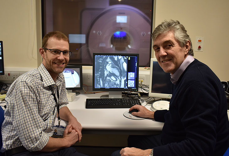
Consultant Cardiologist Dr Andrew Marshall and Consultant Radiologist Dr John Giles viewing the MRI heart scan
An MRI heart scan is used to monitor heart disease, evaluate the heart’s anatomy and function investigating the blood supply to the heart, heart muscle conditions, damage to the heart muscle and heart valve disease.
Professor Nik Patel, Consultant Cardiologist said: “This new scan will be a significant addition to our range of cardiac investigations. It will mean that fewer patients will need to travel to specialist hospitals in London for this test.”
Dr Justin Harris, Consultant Radiologist said: “The new MRI scanner will help us build on our cardiac imaging service. It means we are able to undertake more detailed investigations, improving our diagnostic capability.”
Magnetic resonance imaging (MRI) uses a powerful magnetic field, radio waves and a computer to produce detailed pictures of the structures within and around the heart. MRI scans do not use ionizing radiation to produce images providing the best images of the heart for certain conditions.